

Game changer: New 7T MRI elevates Auburn research
September 12, 2024
Cookie Acknowledgement
This website uses cookies to collect information to improve your browsing experience. Please review our Privacy Statement for more information.
The new 7T MRI will allow Auburn to leverage its existing expertise in engineering, sciences and veterinary medicine to improve health care.
Gopikrishna Deshpande wants to see the brain in action.
How does information between billions of neurons flow within this cerebral superhighway? What functional details impact decision-making or even clinical disorders such as autism or Alzheimer’s?
Now, he can.
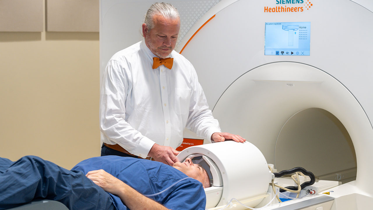
The recently approved $9 million Siemens MAGNETOM Terra.X 7 Tesla (7T) magnetic resonance imaging (MRI) scanner inside the Auburn University Neuroimaging Center takes researchers to places they’ve never been and provides them with detailed, ultra-high-resolution images they previously could not study. The world’s first field-installed, clinically approved parallel transmit 7T MRI scanner complements the center’s 3T scanner, part of its original equipment that’s still used today.
“3T scanners are standard in clinical practice today, and you get a fairly good view of what’s happening in the brain — but you miss a lot of the details,” said Deshpande, a professor of electrical and computer engineering, who specializes in signal/image processing. “Because the brain is such a complex organ with billions of neurons and trillions of connections, the devil is in the details. As researchers, we want to have as much detailed depiction, with high-resolution images, as possible.”
For Auburn, installing the powerful whole-body Terra.X 7 Tesla scanner was … a no-brainer.
“The university is sitting on a gold mine in terms of data and capabilities,” said Thomas Denney, director of the Auburn University Neuroimaging Center and the Mr. & Mrs. Bruce Donnellan & Family Endowed Professor in the Department of Electrical and Computer Engineering. “This machine puts us on the map in the MRI universe, and it allows us to play in a sandbox that we couldn’t play in before.”
Denney said updated scanners like this are fitted with parallel transmit technology and sodium imaging capability.
“With our old 7T, if we did an MRI scan of the brain in certain regions, you wouldn’t receive uniform coverage of the brain,” Denney said. “That’s because we had only one transmit channel — like a flashlight inside a large room with only one beam. By comparison, our new Terra.X 7 provides us with eight beams of light that illuminate the brain and provide a more uniform image. Sodium imaging technology is important because it can measure how much sodium is in your brain regionally and identify abnormalities.”
Denney noted that Auburn researchers, many whom have already utilized the center’s previous 7T and 3T equipment, will take advantage of this new avenue. Fresh research explorations already underway include:

Jennifer Robinson’s teaching and research in neuroscience will be elevated with the use of the new 7T MRI scanner on campus.
“I dreamed of building an MRI research program here at Auburn where large medical centers recognize our contributions,” Denney said. “Through research, Auburn University has moved the needle and more than exceeded my expectations. Since opening the Thomas Walter MRI Research Building in 2010, we’ve been able to double our staff. Those machines are great, but people are the ones who write research papers, and people are the ones who earn those research grants.”
People like Deshpande.
“MRI research has come a long way — and it will continue to evolve,” Deshpande said. “I’m proud that Auburn University is a leading player in this field as my colleagues and myself continue to explore the unexplored, explain the unexplained and provide impactful research to improve the quality of life for all.”